

February 17, 2011
Story by Gregory Kennedy
“I came in really, really scared last night, thinking that it’s my last day,” says David Corneau, only hours after brain surgery.
“And I’m going home today to play with my kids."
This 33-year-old Edmonton father of three was able to go home a day after aneurysm surgery thanks to a new microscope that makes blood vessels in the brain light up like neon. 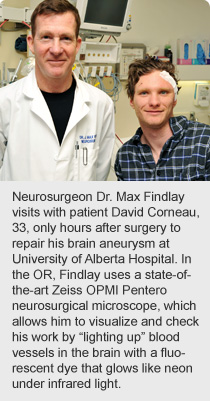
The technology allows neurosurgeons at the University of Alberta Hospital to verify that procedures to restore healthy blood flow are successful while still in the operating room, improving patient outcomes and reducing the need for followup surgeries to eliminate new leaks or blockages in blood vessels.
“We get a picture of the blood vessels that we’ve never had before,” says neurosurgeon Dr. Max Findlay. “The blood vessels shine silvery white against a dark background, which gives us the security and knowledge right in the operating room that an aneurysm is completely gone, obliterated, and all the healthy blood vessels around it are left intact.”
The University of Alberta Hospital is the only facility in Alberta that uses the $380,000 Zeiss OPMI Pentero, which was completely funded by the University Hospital Foundation. It’s the world’s first surgical microscope built specifically to support a procedure called fluorescence angiography. A special green dye is injected intravenously and made visible by infrared light as it flows through the arteries, capillaries and veins of the brain.
About a dozen patients have benefited from the technology since it was introduced at University of Alberta Hospital in late December. Many of these patients have vascular brain conditions such as aneurysm (blister-like bulge that weakens a blood-vessel wall), arteriovenous malformation (abnormal connection between veins and arteries, usually congenital) and fistula (an abnormal connection or passageway between two vessels).
“The beauty of fluorescence angiography is its simplicity. The anesthetist just injects the dye into a peripheral vein and, in seconds, you’re looking through the microscope at the surgical target,” says Findlay.
“Before, we relied on a complicated catheter angiogram. We’d put a catheter in an artery, feed it to the arteries of the brain and inject a dye — not a fluorescent dye — that makes the blood vessels stand out as black on an X-ray. This standard angiogram is still a safe, effective procedure but it typically requires us to keep the patient in hospital for an additional day or two to verify its success. Fluorescence angiography is just better technology.”
David Corneau says he was feeling better, at home, within 24 hours of surgery.
“I completely got my strength back already,” he says. “The technology is pretty cool and the care has been amazing. I had a nurse sit up with me at the end of my bed, talking to me all night long before surgery, answering my questions, pouring me a glass of water. Just being there for me, giving me support. That was nice.”
Community generosity helped to make it all possible.
“We would not be able to have this advanced technology at our hospital right now without the support of the many donors to our annual campaign and the $1 million raised through the Festival of Trees in support of neurosciences,” says Joyce Mallman Law, president of University Hospital Foundation.
For health advice 24/7 phone 811 a free health information service. You have options to speak with a variety of health professionals.
Alberta Health Services values your input. Your feedback will help us continuously improve the quality of Alberta's healthcare system.
Online payment is available for hospital stays, food handling permits, ambulance services, courses, parking citations, and parking permits.